Ytterbium »
PDB 1c5k-3fty »
1ytt »
Ytterbium in PDB 1ytt: Yb Substituted Subtilisin Fragment of Mannose Binding Protein-A (Sub- Mbp-A), Mad Structure at 110K
Protein crystallography data
The structure of Yb Substituted Subtilisin Fragment of Mannose Binding Protein-A (Sub- Mbp-A), Mad Structure at 110K, PDB code: 1ytt
was solved by
F.T.Burling,
W.I.Weis,
K.M.Flaherty,
A.T.Brunger,
with X-Ray Crystallography technique. A brief refinement statistics is given in the table below:
| Resolution Low / High (Å) | 10.00 / 1.80 |
| Space group | P 21 21 21 |
| Cell size a, b, c (Å), α, β, γ (°) | 65.508, 72.216, 45.035, 90.00, 90.00, 90.00 |
| R / Rfree (%) | 18.5 / 20.6 |
Ytterbium Binding Sites:
The binding sites of Ytterbium atom in the Yb Substituted Subtilisin Fragment of Mannose Binding Protein-A (Sub- Mbp-A), Mad Structure at 110K
(pdb code 1ytt). This binding sites where shown within
5.0 Angstroms radius around Ytterbium atom.
In total 4 binding sites of Ytterbium where determined in the Yb Substituted Subtilisin Fragment of Mannose Binding Protein-A (Sub- Mbp-A), Mad Structure at 110K, PDB code: 1ytt:
Jump to Ytterbium binding site number: 1; 2; 3; 4;
In total 4 binding sites of Ytterbium where determined in the Yb Substituted Subtilisin Fragment of Mannose Binding Protein-A (Sub- Mbp-A), Mad Structure at 110K, PDB code: 1ytt:
Jump to Ytterbium binding site number: 1; 2; 3; 4;
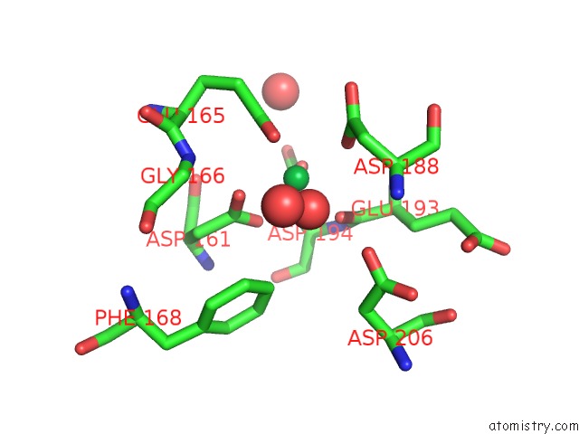
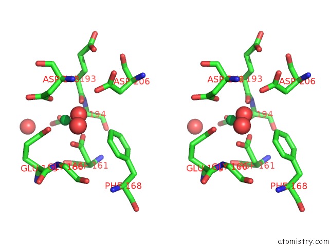
Ytterbium binding site 1 out of 4 in 1ytt
Go back to
Ytterbium binding site 1 out
of 4 in the Yb Substituted Subtilisin Fragment of Mannose Binding Protein-A (Sub- Mbp-A), Mad Structure at 110K
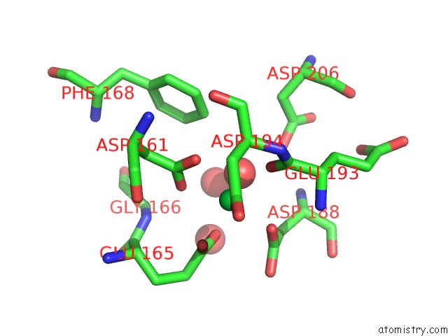
Mono view
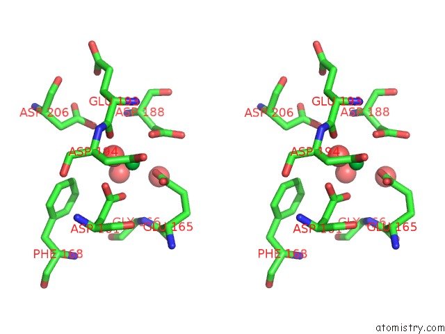
Stereo pair view
Mono view
Stereo pair view
A full contact list of Ytterbium with other atoms in the Yb binding
site number 1 of Yb Substituted Subtilisin Fragment of Mannose Binding Protein-A (Sub- Mbp-A), Mad Structure at 110K within 5.0Å range:
|
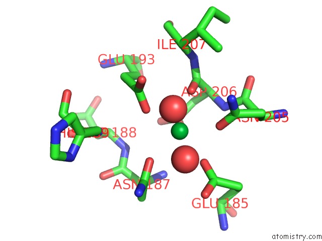
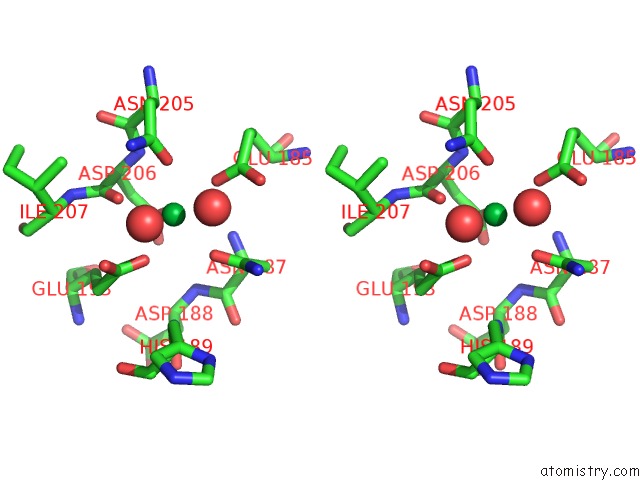
Ytterbium binding site 2 out of 4 in 1ytt
Go back to
Ytterbium binding site 2 out
of 4 in the Yb Substituted Subtilisin Fragment of Mannose Binding Protein-A (Sub- Mbp-A), Mad Structure at 110K
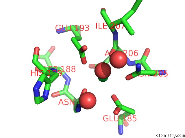
Mono view
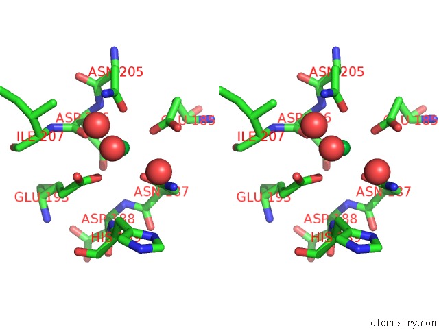
Stereo pair view
Mono view
Stereo pair view
A full contact list of Ytterbium with other atoms in the Yb binding
site number 2 of Yb Substituted Subtilisin Fragment of Mannose Binding Protein-A (Sub- Mbp-A), Mad Structure at 110K within 5.0Å range:
|
Ytterbium binding site 3 out of 4 in 1ytt
Go back to
Ytterbium binding site 3 out
of 4 in the Yb Substituted Subtilisin Fragment of Mannose Binding Protein-A (Sub- Mbp-A), Mad Structure at 110K
Mono view
Stereo pair view
Mono view
Stereo pair view
A full contact list of Ytterbium with other atoms in the Yb binding
site number 3 of Yb Substituted Subtilisin Fragment of Mannose Binding Protein-A (Sub- Mbp-A), Mad Structure at 110K within 5.0Å range:
|
Ytterbium binding site 4 out of 4 in 1ytt
Go back to
Ytterbium binding site 4 out
of 4 in the Yb Substituted Subtilisin Fragment of Mannose Binding Protein-A (Sub- Mbp-A), Mad Structure at 110K
Mono view
Stereo pair view
Mono view
Stereo pair view
A full contact list of Ytterbium with other atoms in the Yb binding
site number 4 of Yb Substituted Subtilisin Fragment of Mannose Binding Protein-A (Sub- Mbp-A), Mad Structure at 110K within 5.0Å range:
|
Reference:
F.T.Burling,
W.I.Weis,
K.M.Flaherty,
A.T.Brunger.
Direct Observation of Protein Solvation and Discrete Disorder with Experimental Crystallographic Phases. Science V. 271 72 1996.
ISSN: ISSN 0036-8075
PubMed: 8539602
Page generated: Sat Oct 12 20:57:57 2024
ISSN: ISSN 0036-8075
PubMed: 8539602
Last articles
F in 7JXMF in 7JUY
F in 7JV0
F in 7JUU
F in 7JV1
F in 7JUZ
F in 7JUT
F in 7JUV
F in 7JUX
F in 7JH6