Ytterbium »
PDB 1c5k-3fty »
1ytt »
Ytterbium in PDB 1ytt: Yb Substituted Subtilisin Fragment of Mannose Binding Protein-A (Sub- Mbp-A), Mad Structure at 110K
Protein crystallography data
The structure of Yb Substituted Subtilisin Fragment of Mannose Binding Protein-A (Sub- Mbp-A), Mad Structure at 110K, PDB code: 1ytt
was solved by
F.T.Burling,
W.I.Weis,
K.M.Flaherty,
A.T.Brunger,
with X-Ray Crystallography technique. A brief refinement statistics is given in the table below:
| Resolution Low / High (Å) | 10.00 / 1.80 |
| Space group | P 21 21 21 |
| Cell size a, b, c (Å), α, β, γ (°) | 65.508, 72.216, 45.035, 90.00, 90.00, 90.00 |
| R / Rfree (%) | 18.5 / 20.6 |
Ytterbium Binding Sites:
The binding sites of Ytterbium atom in the Yb Substituted Subtilisin Fragment of Mannose Binding Protein-A (Sub- Mbp-A), Mad Structure at 110K
(pdb code 1ytt). This binding sites where shown within
5.0 Angstroms radius around Ytterbium atom.
In total 4 binding sites of Ytterbium where determined in the Yb Substituted Subtilisin Fragment of Mannose Binding Protein-A (Sub- Mbp-A), Mad Structure at 110K, PDB code: 1ytt:
Jump to Ytterbium binding site number: 1; 2; 3; 4;
In total 4 binding sites of Ytterbium where determined in the Yb Substituted Subtilisin Fragment of Mannose Binding Protein-A (Sub- Mbp-A), Mad Structure at 110K, PDB code: 1ytt:
Jump to Ytterbium binding site number: 1; 2; 3; 4;
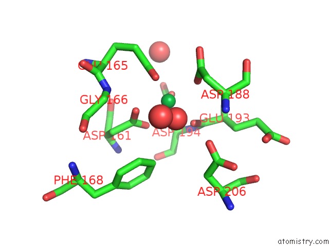
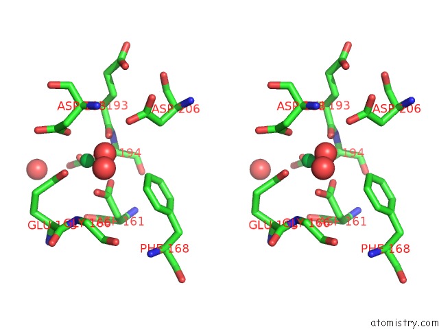
Ytterbium binding site 1 out of 4 in 1ytt
Go back to
Ytterbium binding site 1 out
of 4 in the Yb Substituted Subtilisin Fragment of Mannose Binding Protein-A (Sub- Mbp-A), Mad Structure at 110K
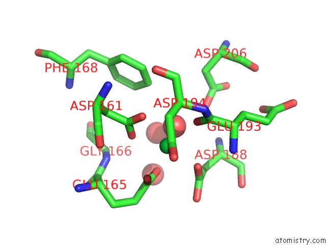
Mono view
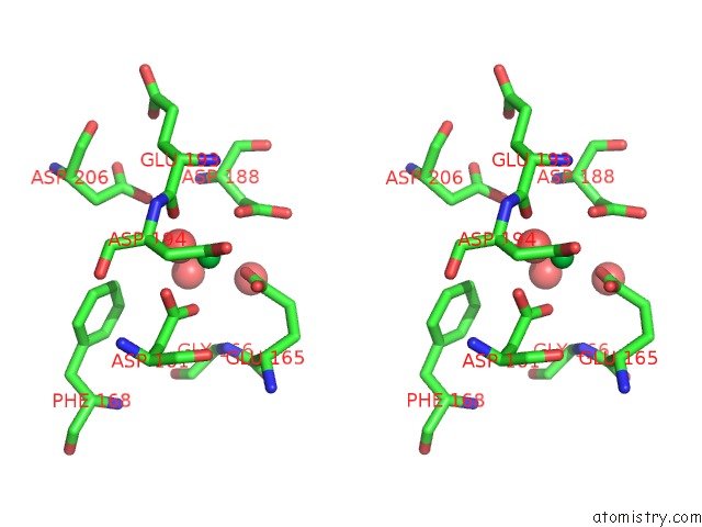
Stereo pair view
Mono view
Stereo pair view
A full contact list of Ytterbium with other atoms in the Yb binding
site number 1 of Yb Substituted Subtilisin Fragment of Mannose Binding Protein-A (Sub- Mbp-A), Mad Structure at 110K within 5.0Å range:
|
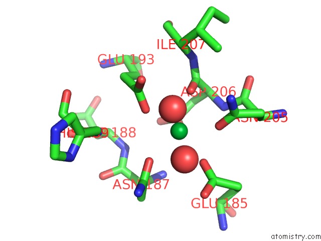
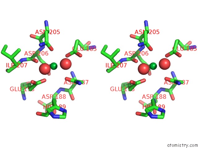
Ytterbium binding site 2 out of 4 in 1ytt
Go back to
Ytterbium binding site 2 out
of 4 in the Yb Substituted Subtilisin Fragment of Mannose Binding Protein-A (Sub- Mbp-A), Mad Structure at 110K
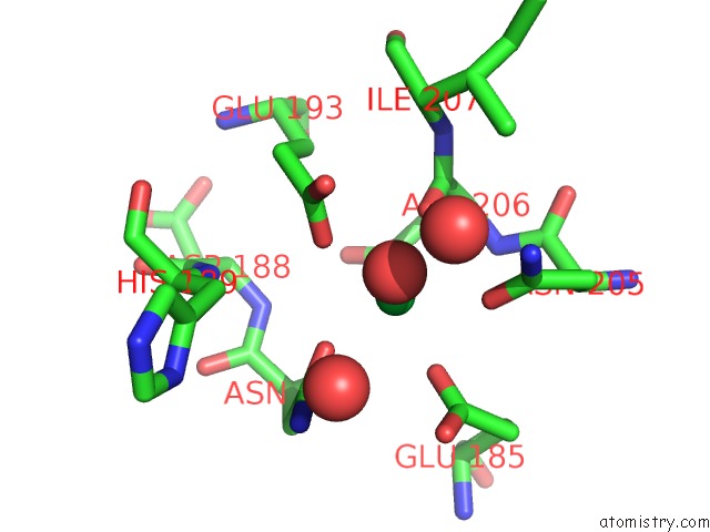
Mono view
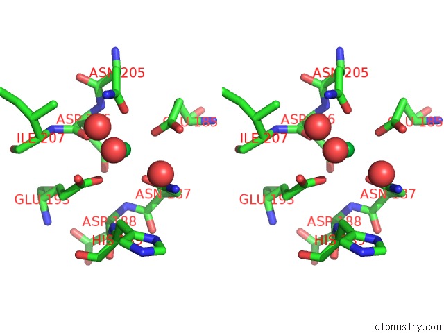
Stereo pair view
Mono view
Stereo pair view
A full contact list of Ytterbium with other atoms in the Yb binding
site number 2 of Yb Substituted Subtilisin Fragment of Mannose Binding Protein-A (Sub- Mbp-A), Mad Structure at 110K within 5.0Å range:
|
Ytterbium binding site 3 out of 4 in 1ytt
Go back to
Ytterbium binding site 3 out
of 4 in the Yb Substituted Subtilisin Fragment of Mannose Binding Protein-A (Sub- Mbp-A), Mad Structure at 110K
Mono view
Stereo pair view
Mono view
Stereo pair view
A full contact list of Ytterbium with other atoms in the Yb binding
site number 3 of Yb Substituted Subtilisin Fragment of Mannose Binding Protein-A (Sub- Mbp-A), Mad Structure at 110K within 5.0Å range:
|
Ytterbium binding site 4 out of 4 in 1ytt
Go back to
Ytterbium binding site 4 out
of 4 in the Yb Substituted Subtilisin Fragment of Mannose Binding Protein-A (Sub- Mbp-A), Mad Structure at 110K
Mono view
Stereo pair view
Mono view
Stereo pair view
A full contact list of Ytterbium with other atoms in the Yb binding
site number 4 of Yb Substituted Subtilisin Fragment of Mannose Binding Protein-A (Sub- Mbp-A), Mad Structure at 110K within 5.0Å range:
|
Reference:
F.T.Burling,
W.I.Weis,
K.M.Flaherty,
A.T.Brunger.
Direct Observation of Protein Solvation and Discrete Disorder with Experimental Crystallographic Phases. Science V. 271 72 1996.
ISSN: ISSN 0036-8075
PubMed: 8539602
Page generated: Sat Oct 12 20:57:57 2024
ISSN: ISSN 0036-8075
PubMed: 8539602
Last articles
Na in 1VQ7Na in 1VQ5
Na in 1VQ6
Na in 1VQ4
Na in 1VI6
Na in 1VKG
Na in 1VMJ
Na in 1VMH
Na in 1VMF
Na in 1VLM